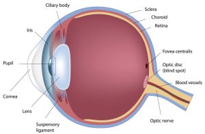
The eye is one of the sense organs of the body. It is made of different parts that have different functions but work together to enable us see. It is like a ball, with most of it sitting in a hole in the skull known as the eye socket or orbit.
It has a multilayered wall, a space inside it that contains the lens and the fluids that help to keep it in shape and other parts known as accessory structures.
The eye, in its orbit, is surrounded by orbital fats and fibrous tissues that cushion it, as well as extraocular muscles that move it in different directions and in front, it is protected by the eyelids, conjunctiva and tears from the lacrimal system.
The wall of the eye
The wall of the eye consists of three layers namely:
- The outer layer, which consists of the cornea and sclera
- The middle layer, which consists of the iris, the ciliary body and the choroid
- The inner layer, which consists of the retina
The outer layer
This is made up of thick fibrous tissues that protect the inner structures of the eye. The outer layer consists of the cornea and sclera.
Cornea: This is the transparent structure at the front of the eye. It is located in front of the pupil. Its major function is to bend light rays entering the eye to enable them to focus on the retina. Therefore, it is the main refractive surface of the eye and provides most of its focusing power.
Sclera: This is the white coat of the eye. It is the toughest structure of the eye and serves to protect the eye from penetrating injuries. It helps in maintaining the shape of the eyeball together with the fluid in the eye. It also provides attachments for the muscles that control eye movements.
The middle layer
The middle layer is the vascular part of the eye. It is known as the uvea and consists of the iris, ciliary body and choroid.
Iris: This is the coloured part of the eye. It has a rich supply of blood vessels and contains muscles that control the size of the pupil (the opening in the iris through which light enters the eye). One of the muscles known as the dilator muscle causes the pupil to widen in dim light. Another muscle known as the sphincter muscle, causes the pupil to constrict in bright light. The amount of pigment in the iris determines the colour of the eye. The major function of the iris is to control the amount of light entering the eye.
Ciliary body:The ciliary body is part of the middle layer of the eye connecting the iris to the choroid. It is attached to the crystalline lens by ligaments and holds the lens in place behind the pupil. It contains muscles that control the ability of the lens to change its focusing power at different distances (a process known as accommodation). It also produces aqueous humour (the fluid that fills the anterior chamber of the eye and maintains intraocular pressure).
Choroid: The choroid is the posterior part of the middle layer of the eye. It is a connective tissue richly supplied with blood vessels and nerves. Its major function is to provide nutrition for the retina and the sclera.
Inner layer
The inner layer of the eye is the most sensitive part of the eye known as the retina.
Retina: The retina is the most sensitive part of the eye. It is responsible for visual perception. It consists of nervous tissues with multiple sensitive cells that all work together under normal conditions to produce good vision. On the retina, there are two major light-sensitive cells (photoreceptors) responsible for vision. These are the rods and cones. The rods are responsible for night vision while the cones are responsible for coloured daylight vision. Sensations received by these photoreceptors are transmitted through the optic nerve (a cranial nerve) to the brain where they are interpreted.
The inner part of the eye
The inner part of the eye is the area surrounded by a three-layered wall. It contains the lens and spaces (chambers) filled with aqueous and vitreous humour.
Lens: The lens is a transparent oval-shaped structure in the eye. It is held in place behind the people by ligaments attached to the ciliary body. It focuses light rays entering the eye on the retina.
Aqueous humour: Aqueous humour is one of the fluids in the eye. It is produced by the ciliary body and fills the spaces in the anterior part of the eye; that is the space between the cornea and the iris (anterior chamber) and that between the iris and the lens (posterior chamber). Aqueous humour provides nutrients for the cornea and lens and helps to keep the eye in shape.
Vitreous Humour: The vitreous humour is the fluid that fills the space between the lens and retina, known as the vitreous chamber. It protects the retina and keeps it in place. It also helps the eye maintain its shape.
Accessory structures
These are structures external to the eyeball that work with other components of the eye to ensure good vision. They include the conjunctiva, eyelid, lacrimal system and extraocular muscles.
Conjunctiva: This is a thin layer of transparent tissue that covers the sclera and lines the inner surface of the eyelid. It lubricates the eye and serves as a protective barrier for the front surface of the eye.
Eyelid: The eyelid is the fold of skin that covers the eye and protects it from environmental hazards and injury.
Lacrimal System: This consists mainly of the lacrimal gland, lacrimal sac and nasolacrimal duct which respectively produces, reserves and drains the tears that lubricate the eyes.
Extraocular Muscles: These are 6 muscles within the orbit that are attached to the outer layer of the wall of the eye at different positions. They keep the eye in place and are responsible for moving the eye in different directions.